News
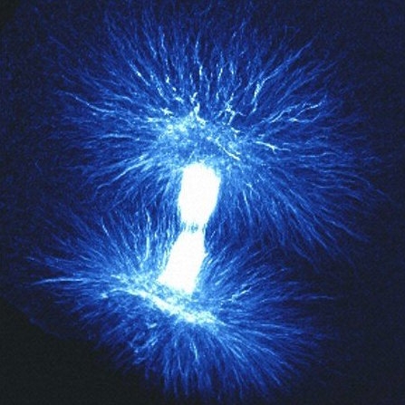
The Needleman Lab developed a model describing how spindles (above) assemble to play their vital role in cell division, separating chromosomes and pulling the duplicated DNA from the mother cell into the daughter cell. (Image courtesy of B. Strauss/Cell Image Library)
What bones are to bodies, the cytoskeleton is to cells. The cytoskeleton maintains cellular structure, builds appendages like flagella and, together with motor proteins, powers cellular movement, transport, and division. Microtubules are a critical component of the cytoskeleton, vital for cell division and, because of that, an excellent target for chemotherapy drugs.
Microtubules can spontaneously self-organize, transforming from many singular components into one large cellular structure capable of performing specific tasks. Think Transformers. How they do that, however, has remained unclear.
Now, researchers at the Harvard John A. Paulson School of Engineering and Applied Sciences (SEAS) have observed how microtubules and motor proteins assemble into macroscopic networks. Their observation provides a better understanding of cytoskeletal self-organization in general, which may in turn lead to better drug design and new materials that can mimic cellular behaviors.
The research was recently published in the journal eLife.
Spindles are cellular structures that play an important role in cell division, separating chromosomes and pulling the duplicated DNA from the mother cell into the daughter cell. They are made up of microtubules and many other proteins, including the motor protein dynein.
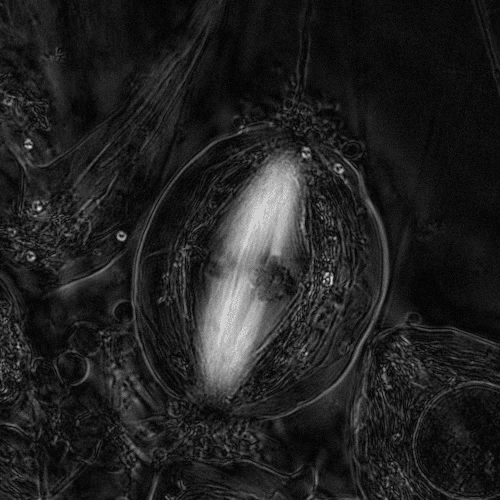
The white spindles in the center of the cell separate the chromosomes and pull the duplicated DNA from the mother cell into the daughter cell(Video courtesy of Rudolf Oldenbourg and James R. LaFountain/Cell Image Library)
“What we are really looking for is a grand unified theory of spindle assembly,” said Peter Foster, a graduate student at SEAS and the paper’s first author. “We know how motor proteins interact with microtubules but how do you go from individual microtubules and motor proteins to large networked structures?”
To gain insight into how spindles assemble, Foster and his team, under the leadership of Dan Needleman, associate professor of applied physics and of molecular and cellular biology, built a simple experiment. They extracted cytoplasm from frog eggs, which contains dynein and all of the components needed to make spindles, added fluorescent protein and the chemotherapy drug Taxol to create and stabilize microtubules, and loaded the mixture into “the world’s simplest microfluidic chamber.”
“Very quickly, we saw that these microtubules organize into networks that spontaneously contract,” Foster said. “The question is why?”
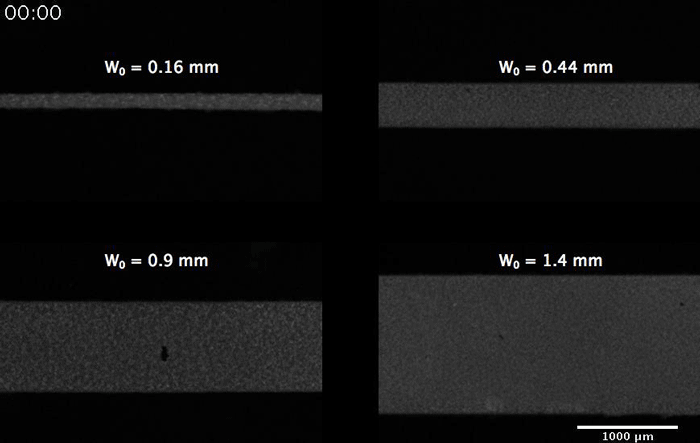
The microtubles organize into networks that spontaneously contract, regardless of the width of the chamber (Image Needleman Lab/HarvardSEAS)
The answer lay not in the microtubules but in the behavior of the motor protein. Microtubules have plus and minus ends and researchers have observed dynein moving from the plus end to the minus. As a result, the motor protein draws the minus ends of microtubules together, creating star-like clusters called asters. The dynein drives these small clusters together, fusing them to create larger and larger networks. As the motor protein continues to jam the microtubules together, the network contracts, until it can’t get any smaller.
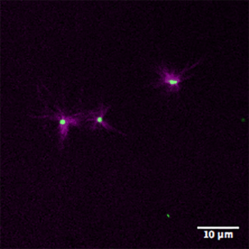
Asters form as motor protein draws the minus ends of the purple microtubules together (Image Needleman Lab/Harvard SEAS)
Based on this experiment, the researchers developed a model that quantifies and describes this behavior and lends insight into not only spindle assembly but also self-organization in general. This model could provide insights into how to design materials that can self-assemble or autonomously contract, like a self-squeezing sponge.
“Using this model, we can ask questions from the microscopic level all the way to large scale phenomena,” Foster said. “There are many ramifications not only in biology but also in the material world."
This paper was co-authored by Sebastian Fürthauer and Michael Shelley. The research was supported by the National Science Foundation, the National Institutes of Health, and the Human Frontier Science Program.
Topics: Materials
Cutting-edge science delivered direct to your inbox.
Join the Harvard SEAS mailing list.
Press Contact
Leah Burrows | 617-496-1351 | lburrows@seas.harvard.edu
Related News


3 tech solutions to societal needs will get help moving to market
Harvard Office of Technology Development
Applied Physics, Bioengineering, Health / Medicine, Materials, Materials Science & Mechanical Engineering, Optics / Photonics, Quantum Engineering