News
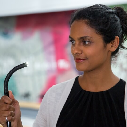
The antifouling surface coating for endoscopes, developed at the Wyss Institute by Core Faculty member Joanna Aizenberg and her team, drastically improves camera-guided field of vision and reduces procedure time. Steffi Sunny, co-first author of the study recently published in PNAS, is pictured holding an endoscope. (Image courtesy of Wyss Institute at Harvard University)
With the help of camera-guided endoscopes, clinicians get a look inside the body’s cavities to diagnose and treat many different conditions. In the United States alone, up to 20 million endoscopies are performed on patients every year. But even for the most seasoned endoscope users, the instruments can prove very challenging to use effectively; this is due to the fact that blood and other bodily fluids quite commonly obscure the camera lens in the midst of critical procedures.
This problem inspired a team led by Joanna Aizenberg, Amy Smith Berylson Professor of Materials Science and Professor of Chemistry & Chemical Biology at the Harvard John A. Paulson School of Engineering and Applied Sciences (SEAS) and Core Faculty member of the Wyss Institute for Biologically Inspired Engineering at Harvard University, to engineer a transparent surface coating for an endoscope lens that could effortlessly keep blood and other fluids at bay. The idea took roots in conversations Aizenberg had with clinician collaborators, who were quick to lament the propensity for endoscopes to become clouded mid-procedure.
Turning to the portfolio of SLIPS (Slippery Liquid-Infused Porous Surfaces) technologies already invented by Aizenberg, they set out to design a specialized SLIPS coating that could prevent bodily fluids from blocking the optical field of view of camera-guided endoscopes. The demonstrated results of their work are published online this week in the Proceedings of the National Academy of Sciences (PNAS) journal.
“Endoscopes are used by many physicians around the world for a variety of procedures, and ironically, the moment when bleeding occurs and the optical field is blocked is also precisely when physicians most need to see what’s happening,” said Aizenberg, who is also the Co-Director of the Kavli Institute for Bionano Science and Technology.
Aizenberg’s SLIPS technology creates non-wetting and robust self-cleaning surfaces that can resist almost any fouling challenge a surface may face. But to develop the technology for endoscopic use, the team needed to adapt SLIPS specifically to weather the harsh environment of a living body’s cavities.
“In addition to being entirely transparent and able to coat the curvature of the glass camera lens on the endoscope, the coating also needs to withstand constant contact and abrasion with soft tissues and corrosive bodily fluids,” said Steffi Sunny, a co-first author on the study and a graduate student at SEAS and researcher at the Wyss.
To achieve this, the team deposited silica nanoparticles layer by layer onto an endoscope’s glass camera lens. These silica layers create a porous surface that, at the nanoscale, would be considered “rough” and filled with caverns. They then functionalized this “rough” surface and infused it with a medical-grade silicone oil, filling in the porous cavities and creating a self-replenishing liquid layer. The end result, an entirely biocompatible coating, can endure many procedural uses and standard sterilization protocols, and can be even re-applied with silicone oil intermittently to maintain its extreme repellency.
Working with co-first author George Cheng the team tested their antifouling endoscope in vivo, specifically in bronchoscopy. Bronchoscopy is one of the most commonly performed procedures on patients with pathological lung conditions. Cheng, who was an Interventional Pulmonary Fellow at Harvard Combined Training Program (Beth Israel Deaconess Medical Center and Massachusetts General Hospital) at the time of the study, performed bronchoscopy with the modified endoscope on an anesthetized pig to evaluate its effectiveness.
“The proof of concept experiment in the pig lung worked beautifully,” said Cheng, who today is Assistant Professor of Medicine in the Division of Pulmonary, Allergy and Critical Care at Duke University. “It very easily repelled blood and mucus and dramatically reduced the complexity of the procedure.”
Conventional scopes generally cause the procedure time to be longer than necessary, due to the need to repeatedly clean and wipe away fluids that obscure the optical field. An antifouling endoscope could, one day, not only improve ease and precision of endoscopies, but could also result in more positive patient outcomes due to a shortened procedural time, significantly reduced side-effects often associated with mid-procedure lens cleaning, less sedation needed, and decreased recovery time.
SLIPS-coated endoscopes could even expand our view into new areas of the body that we have not yet been able to access with a camera. Today’s endoscopes, which contain irrigation and suction channels to flush away build up from the lens mid-procedure, are limited in how small they can be made due to this important cleansing feature. But the super repellent coating on the lens could potentially eliminate the need for a wash port, leading to more miniaturized endoscopes, which would allow physicians to reach, observe and treat the areas of the body that are off-limits to endoscopes of current sizes.
Looking ahead, the team also hopes that their slippery lens coating could have applications in other camera-guided instruments that operate in harsh, contaminated environments and rely on an unobstructed optical field, such as oil exploration, robotics, marine exploration, plumbing and sanitation and drain pipe maintenance.
In addition to Aizenberg, Sunny, and Cheng, other authors on the new study include: Daniel Daniel (SEAS); Peter Lo (Interventional Pulmonary at BIDMC); Sebastian Ochoa (Interventional Pulmonary at BIDMC); Caitlin Howell (SEAS, University of Maine – Orono, Wyss Institute); Nicolas Vogel (Institute of Particle Technology and Interdisciplinary Center for Functional Particle Systems at Friedrich-Alexander University Erlangen-Nürnberg); and Adnan Majid (Interventional Pulmonary at BIDMC).
Topics: Materials
Cutting-edge science delivered direct to your inbox.
Join the Harvard SEAS mailing list.
Scientist Profiles
Joanna Aizenberg
Amy Smith Berylson Professor of Materials Science and Professor of Chemistry & Chemical Biology
Press Contact
Leah Burrows | 617-496-1351 | lburrows@seas.harvard.edu


