News
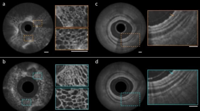
Comparison of images of fruit flesh (left) and swine airways (right) obtained using the nano-optic endoscope (b,d) and a conventional catheter (a,c). (Credit: Harvard University/Massachusetts General Hospital)
The diagnosis of diseases based in internal organs often relies on biopsy samples collected from affected regions. But collecting such samples is highly error-prone due to the inability of current endoscopic imaging techniques to accurately visualize sites of disease. The conventional optical elements in catheters used to access hard-to-reach areas of the body, such as the gastrointestinal tract and pulmonary airways, are prone to aberrations that obstruct the full capabilities of optical imaging.
Now, experts in endoscopic imaging at Massachusetts General Hospital (MGH) and pioneers of flat metalens technology at the Harvard John A. Paulson School of Engineering and Applied Sciences (SEAS), have teamed up to develop a new class of endoscopic imaging catheters – termed nano-optic endoscopes – that overcome the limitations of current systems.
The research is described in Nature Photonics.
“Clinical adoption of many cutting-edge endoscopic microscopy modalities has been hampered due to the difficulty of designing miniature catheters that achieve the same image quality as bulky desktop microscopes,” said Melissa Suter, an assistant professor of Medicine at MGH and Harvard Medical School (HMS) and co-senior author of the paper. “The use of nano-optic catheters that incorporate metalenses into their design will likely change the landscape of optical catheter design, resulting in a dramatic increase in the quality, resolution, and functionality of endoscopic microscopy. This will ultimately increase clinical utility by enabling more sophisticated assessment of cell and tissue microstructure in living patients.”
“Metalenses based on flat optics are a game changing new technology because the control of image distortions necessary for high resolution imaging is straightforward compared to conventional optics, which requires multiple complex shaped lenses,” said Federico Capasso, the Robert L. Wallace Professor of Applied Physics and Vinton Hayes Senior Research Fellow in Electrical Engineering at SEAS and co-senior author of the paper. “I am confident that this will lead to a new class of optical systems and instruments with a broad range of applications in many areas of science and technology.”
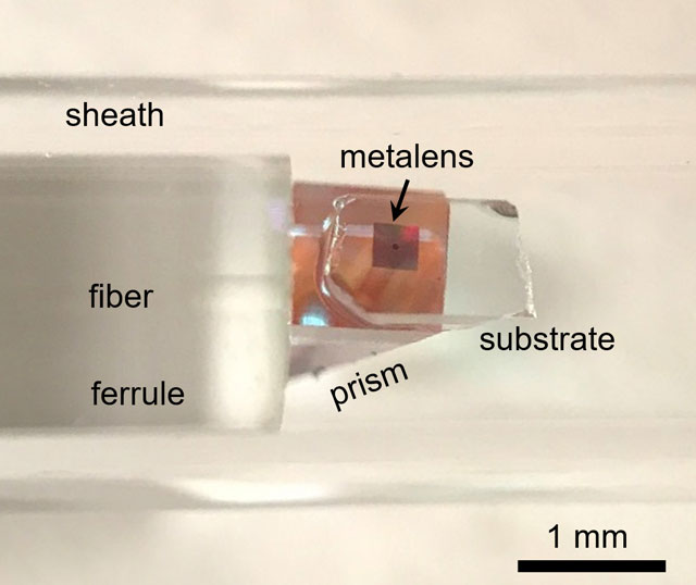
Photographic image of the distal end of the nano-optic endoscope (Credit: Harvard University/Massachusetts General Hospital)
“The versatility and design flexibility of the nano-optic endoscope significantly elevates endoscopic imaging capabilities and will likely impact diagnostic imaging of internal organs,” said Hamid Pahlevaninezhad, Instructor in Medicine at MGH and HMS and co-first author of the paper. “We demonstrated an example of such capabilities to achieve high-resolution imaging at greatly extended depth of focus.”
To demonstrate the imaging quality of the nano-optic endoscope, the researchers imaged fruit flesh, swine and sheep airways, and human lung tissue. The team showed that the nano-optic endoscope can image deep into the tissue with significantly higher resolution than provided by current imaging catheter designs.
The images captured by the nano-optic endoscope clearly show cellular structures in fruit flesh and tissue layers and fine glands in the bronchial mucosa of swine and sheep. In the human lung tissue, the researchers were able to clearly identify structures that correspond to fine, irregular glands indicating the presence of adenocarcinoma, the most prominent type of lung cancer.
“Currently, we are at the mercy of materials that we have no control over to design high resolution lenses for imaging,” said Yao-Wei Huang, a postdoctoral fellow at SEAS and co-first author of the paper. “The main advantage of the metalens is that we can design and tailor its specifications to overcome spherical aberrations and astigmatism and achieve very fine focus of the light. As a result, we achieve very high resolution with extended depth of field without the need for complex optical components.”
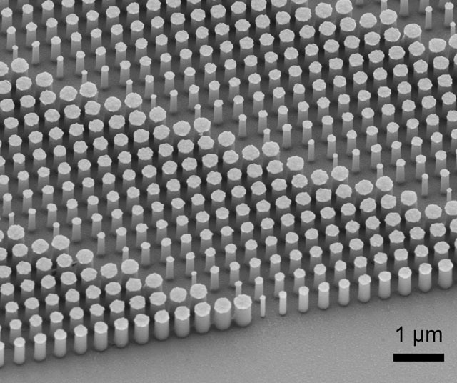
Scanning electron micrograph image of a portion of a fabricated metalens. (Credit: Harvard University/Massachusetts General Hospital)
Next, researchers aim to explore other applications for the nano-optic endoscope, including a polarization-sensitive nano-optic endoscope, which could contrast between tissues that have highly-organized structures, such as smooth muscle, collagen and blood vessels.
This paper was co-authored by Mohammadreza Khorasaninejad, Vivien Ding, and Alexander Zhu, SEAS; Lida P. Hariri and David C. Adams, MGH; Zhujun Shi, Harvard University; and Cheng-Wei Qiu, National University of Singapore.
The research was supported in part by the National Institute of Health, the Air Force Office of Scientific Research and the LUNGevity Foundation/Upstage Lung Cancer.
Topics: Optics / Photonics
Cutting-edge science delivered direct to your inbox.
Join the Harvard SEAS mailing list.
Scientist Profiles

Federico Capasso
Robert L. Wallace Professor of Applied Physics and Vinton Hayes Senior Research Fellow in Electrical Engineering
Press Contact
Leah Burrows | 617-496-1351 | lburrows@seas.harvard.edu


